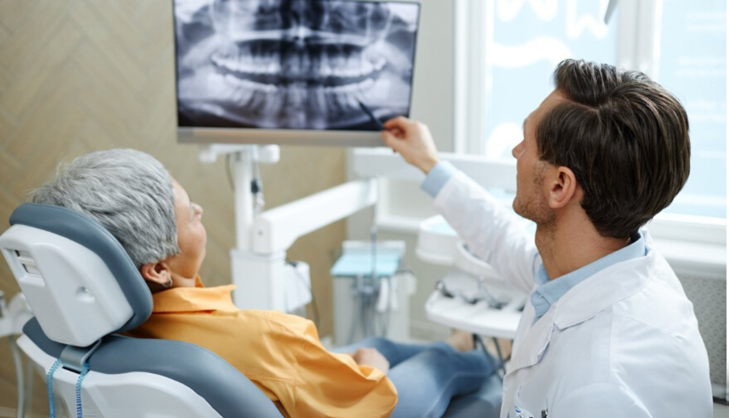
Perhaps in the not-so-distant future, dentists will be able to not only improve oral health, but also simply learn an important indicator – the risk of osteoporosis, thus creating an opportunity to detect this disease, which is often called a silent disease, in time. This could happen thanks to a joint research project* between Riga Stradiņš University (RSU) and the Institute of Electronics and Computer Sciences (EDI).
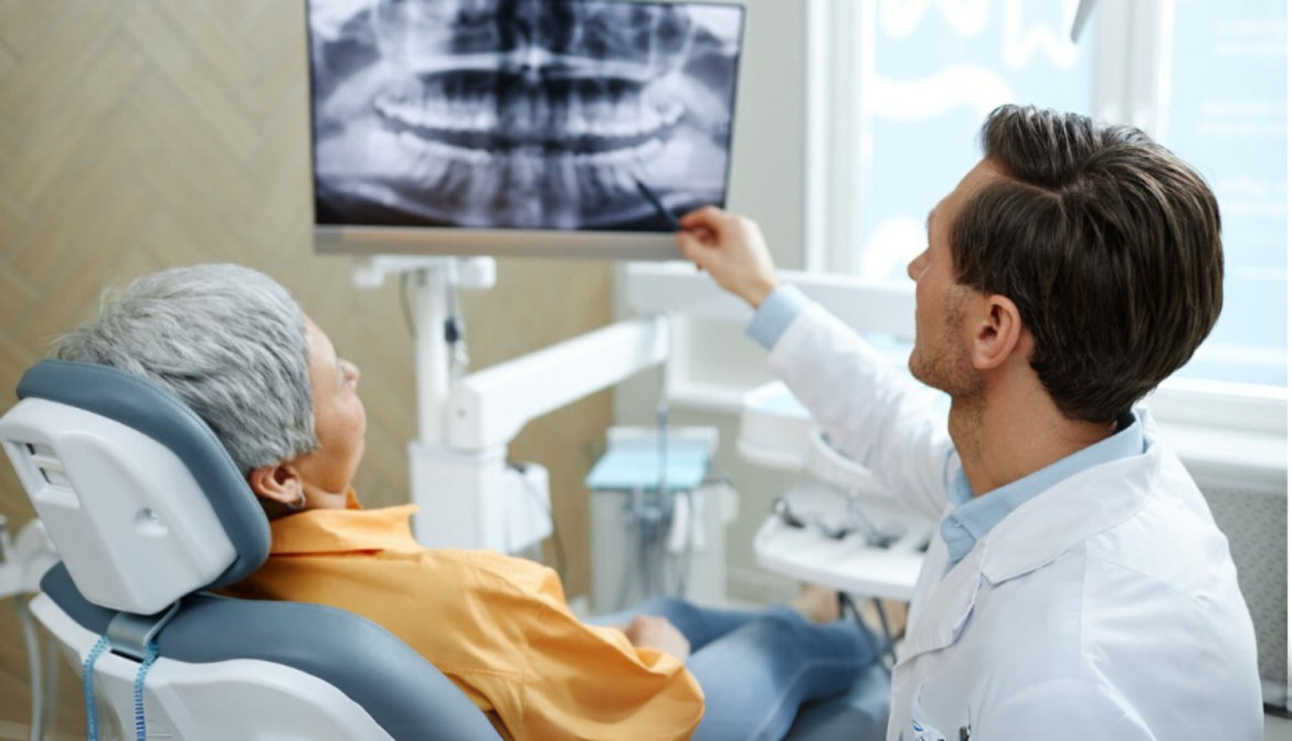
Researchers at RSU and EDI are investigating how cone beam computed tomography examinations, using artificial intelligence (AI), could determine the risk of osteoporosis, selecting patients in the higher risk group, who in turn could undergo further in-depth examinations with a specialised doctor, not a dentist. The head of the research project, Anda Slaidiņa, a leading researcher at the RSU Department of Prosthodontics, tells how the idea began and what we can expect from these studies.
Osteoporosis is often detected late
Osteoporosis is often called a silent disease because there are no pronounced symptoms in the early stages of this skeletal system disease.
"Osteoporosis is a global problem because the population is ageing," says Slaidiņa, continuing: "It is more common in women over the age of 65. Early treatment can prevent serious fractures. In the case of osteoporosis, these can be spinal fractures or hip fractures, which cause disability, sometimes even death. So this disease costs society both financially and emotionally."
The "gold standard" for diagnosing osteoporosis is osteodensitometry, a method for determining bone density. It is an X-ray diagnostic method for determining bone mineral density in the vertebrae of the spine and the hip.
"However, this is not a method that could be used to screen all people of a certain age. Neither in Latvia nor elsewhere in Europe are there enough of these expensive devices to be used for screening. In turn, we are studying how to use relatively easily accessible tests, radiographs, together with a special MI tool at the dentist, so that we can see who is at higher risk of osteoporosis. These people could already be targeted for osteodensitometry and begin treatment," explains the RSU lead researcher.
A dental visit with added value
The beginnings of the research idea can be found in Slaidīna's doctoral thesis 15 years ago, when the researcher had a hypothesis – since postmenopausal patients often visit the dentist, where one of the primary examinations is a review X-ray, it could possibly be used to determine the risk of osteoporosis in cases where this X-ray is already performed in the treatment of dental diseases. So, an additional bonus from an existing examination.
Currently, the idea has already been developed into an even more modern approach, when a special AI tool is also being created – currently, RSU and EDI researchers are continuing to develop it. However, the results are already promising.
"Already in the early days, when the first studies on this topic were published (from 1994), it was clear that conventional X-rays can measure the thickness of the cortical bone (the outer layer of bone) in the lower jaw. If this bone is reduced, the risk of osteoporosis can be well determined. Currently, earlier X-rays are rarely used, basically switching to 3D overview X-rays, and in the study, we looked at how we could use them in examinations. We investigated that 3D X-rays are suitable for determining the risk of osteoporosis," says Slaidiņa.
"The problem is that the primary goal of a dentist is to treat teeth, insert implants, and replace teeth. It would seem that there would be great resistance to putting osteoporosis risk assessment on dentists' shoulders - it would be difficult to implement this method in practice. Let's be honest, there would also be a financial interest - what will the doctor gain from it. Therefore, we realised that there should be a more relaxed method. Namely, RSU and EDI together created an AI tool (which is not yet fully ready, we are improving it), so the dentist does not have to measure anything special - when taking an X-ray, this program automatically gives him a risk result - whether it is present/absent, low, medium or high. And then all that remains is to inform the patient, recommend that they contact a family doctor or osteoporosis specialist for further examinations," explains the RSU lead researcher.
AI research is not easy. Good data is needed to train AI. "This means that there must be an expert who has labelled the data, and the results of the examinations, and there must be a lot of data, then the work of the AI tool will be better. In addition, validation is needed that this method works not only in the X-rays of our study, but also in all other X-rays," says the RSU researcher, thanking EDI, which provides the technical part of the study. "We performed the clinical part, selected the X-ray images to be labelled, but computer scientists provide this information to AI," the scientist says about the collaboration.
To improve the accuracy of the tool, specialists continue to improve it and add other indicators to it, such as changes in the bone structure in the lower jaw. Work is underway to develop a multifactorial model, where data such as the patient's age, weight and height will be added.
--
* "RSU internal and RSU with LSPA external consolidation"
No. 5.2.1.1.i.0/2/24/I/CFLA/005 grant "Conic beam computed tomography as a tool for determining osteoporosis risk in postmenopausal women"
No. RSU-ZG-2024/1-0010