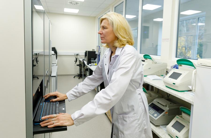
On the day when I interview Professor Aija Linē, she is still the Scientific Director of the Biomedical Research and Study Centre (BMC), as well as leading the cancer biomarker research group at this centre. From June, this work will continue at the National Research and Innovation Institute, in which the BMC is merging with the Institute of Organic Synthesis. In this conversation, I ask how research will continue in the merged institute, as well as about what has already been studied in oncology diagnostics.
Together with colleagues, you have been working for a long time, searching for methods for earlier tumour diagnostics. What are the achievements, and how have the possibilities to diagnose tumours early improved over, for example, the last 10 years?
I would say that evident progress has been achieved relatively recently. For now, however, these developed biomarker tests, with a few exceptions, are not yet widely available and used in the clinic. However, I think that in the fairly near future at least some of the newly developed tests will reach patients. It is not only about Latvia; worldwide as well, blood tests for cancer diagnostics are not yet widely used.
Closer to practical application are the so-called liquid biopsy methods. With their help, it is possible to monitor in the blood how the mutation profile of the tumour changes. This means that cell-free DNA is detected in a blood sample, fragments of which have come from tumour cells and contain mutations of tumour cells. By measuring the amount of these mutations, it is possible to judge whether the disease returns or not in a patient whose tumour has been surgically removed. This provides an opportunity to detect recurrences in a timely manner, as well as to determine whether new mutations have arisen in the cancer cells present in the body.
A liquid biopsy is any analysis in which cancer cells or molecules derived from them are detected in body fluids. Most often these are blood, but it can also be urine, saliva, milk samples, or similar. Thus, in this case, a tissue biopsy is not taken from the tumour, but molecules of cancer cells are detected in a fluid sample. Such tests are already commercially available, therefore they are entering the clinic.
Liquid biopsies have several applications, but they have been most developed in monitoring the effectiveness of anti-cancer therapy. Thus, in patients who have already been diagnosed with a tumour, the liquid biopsy method can be used to monitor tumour changes in the blood or to detect recurrence in a timely manner. Another application could be early diagnostics, but using this method for this purpose would be more difficult, because in this case we do not know exactly what we are looking for in the blood sample. If we know absolutely nothing about a particular patient’s tumour, then sequencing of all cell-free DNA could be performed. In this way, it is possible to attempt to detect changes that could indicate the presence of a tumour.
Speaking about early diagnostics, I know that there are several blood tests currently being tested in clinical trials. However, as far as I know, they are not yet widely used.
Returning to liquid biopsy methods, you yourself are also working on a study in this field. How is it going?
What we are studying are the so-called extracellular vesicles. They are small: from 50 nanometres up to one micrometre. These are vesicles that are enclosed by a cell membrane.
Such vesicles are secreted not only by cells of the human body, but in fact by all forms of life, including bacteria and fungi. Some of the vesicles secreted by cancer cells remain in tumour tissue, while some enter the bloodstream. The vesicles present in the body of a cancer patient contain various molecules that have originated from cancer cells. Therefore, in a human blood sample, we can find vesicles produced by cancer cells, and by analysing either their RNA (ribonucleic acid) or protein composition, we can infer both the presence of a tumour and molecular changes in the tumour. This idea, however, is not very new. From the moment vesicles were discovered, they have been extensively studied.
Vesicles are present in everyone, but it is important that some of the vesicles found in the blood of cancer patients have originated from tumour tissue. Therefore, by taking blood samples and analysing vesicles, it is possible to diagnose a tumour or predict its development. Quite many, including us, have conducted various studies analysing the RNA content in vesicles. We have managed to identify a number of RNA molecules that could serve as biomarkers for breast cancer diagnostics. Most of the markers identified so far have relatively high specificity but low sensitivity. This means that the marker is present only in a small proportion of patients, most often in 10 to 20% of patients. Thus, one marker may be usable only for one tenth of patients. It is, of course, possible to try to combine many such markers into a single test, but the more different markers are included in one test, the more difficult such tests are to validate in the clinic. Therefore, with current technologies, it would be quite difficult to ensure that such tests reach the clinic. This is the conclusion we have reached after 10 years of research.
Similar conclusions have also been reached by other research groups working in this field.
How many years does it take from a scientist’s discovery until this discovery is used in cancer diagnostics and treatment?
Definitely more than 10 years. I would like to say that within the next few years we will see at least the first molecular tests for cancer monitoring.
Would these be available elsewhere in the world or also in Latvia? And to what extent, within the framework of the existing funding, is it possible for medical institutions in Latvia to use the latest medicines and methods?
Funding, of course, is a major problem, but there are also positive examples where it is possible to use what has been developed relatively recently. For example, a couple of years ago the Oncotype test also became available in Latvia. With its help, gene activity in tumour tissue is analysed in breast cancer patients whose tumour has been surgically removed at stage one or two. By determining whether the patient has a high risk that the disease will return, the test helps to decide whether the specific patient needs chemotherapy, which has many unpleasant side effects. If the risk is low, chemotherapy can be avoided. This test has been widely used worldwide for more than ten years. There was a time when it was not available in Latvia, but now it is.
A few years ago, you already discovered that physical activity helps to fight breast cancer. Are studies also being conducted on the impact of an active lifestyle on other types of cancer?
Most likely yes, but we ourselves have not had the opportunity to continue this topic. At present, I am trying to apply for the next projects in this field in order to obtain funding. Our goal is to gain a deeper understanding of the molecular and cellular processes that take place in the human body during physical exertion and to determine how these processes influence tumour behaviour.
Our first study was successful: breast cancer patients receiving neoadjuvant chemotherapy were enrolled in the study. This is chemotherapy administered in courses for about half a year in order to reduce the tumour and make it operable. The patients involved in the study trained regularly—two to three times a week—following specially developed, personalized training programs. First, we observed that in patients who exercised, the effectiveness of chemotherapy was higher than in the control group. Second, by analysing tumour tissues, we found that in patients who exercised, the cellular composition and gene activity in the tumour tissue differed from the control group—those patients who did not change their daily habits during chemotherapy.
The training group had a significantly altered composition of immune cells and immune cell activity. Our hypothesis regarding the interaction between physical activity and the tumour is that during each training session, immune cells that are capable of being activated and migrating enter the bloodstream from various tissue reservoirs, such as the spleen or bone marrow. It had already been shown beforehand that within twenty minutes from the start of exercise, for example, the proportion of NK cells in the blood can increase up to tenfold. After that, these cells can infiltrate the tumour and destroy cancer cells there. Thus, during each training session, the amount of activated immune cells capable of fighting cancer increases slightly in the tumour.
This probably occurs due to the interaction of various factors, as both blood flow increases and the concentrations of different hormones, neurotransmitters, and cytokines in the blood rise. For example, interleukin-6 and adrenaline play an important role. This is the hypothesis that we would like to further test and verify. The benefit for patients would be that, based on these data, we could develop a more precise training program that would better stimulate exactly those processes that are favourable for tumour destruction.
Unfortunately, there is not enough funding even for high-quality and necessary scientific projects. But shouldn’t projects related to health and the relatively large number of oncology patients be a priority?
It is quite a normal process not to obtain funding. In the Latvian Science Council, the competition is such that fewer than one in ten projects receive funding. It is enough for a reviewer to find even a single shortcoming to emphasize for a project not to be approved.
And in my opinion, there is no basis to say that some projects are more of a priority than others. That is precisely why it is an open competition, in which everyone competes under equal conditions. However, it is also true that in any competition there is a certain element of chance, and luck also plays a role.
However, other studies are continuing, for example in the already mentioned field of extracellular vesicles. We are studying a conceptually new technology, the principle of which is based not on the molecular content of vesicles, but rather on how they affect specially engineered biosensor cells. In the study, we take a blood sample from a patient or a healthy individual, isolate vesicles, add them to biosensor cells, and observe how gene activity changes. We expect that vesicles present in the blood of cancer patients will affect processes that are not affected by vesicles from a healthy person.
If this hypothesis is confirmed, over time a fundamentally new type of liquid biopsy tests could enter practice, based not on the detection of molecules of cancer cells, but on their functions.
When was this study started and how far have you progressed in this research?
This is a BioPhot platform project, where the principle is that each project does not last longer than one year. Within this one year, it must be possible to increase the technology readiness level by one stage. If, when submitting the project, we were at TRL 2, then within one year we have to reach TRL 3. If we succeed, we will be able to apply in the next round with the hope of receiving funding for the next steps. Thus, within one year it is necessary to take one significant step.
TRL is a set of terms that describe the technology readiness level. TRL 1 is only a hypothesis. TRL 2 means that there is already evidence for this technology, but nothing has yet been validated in practice. At TRL 3, researchers are already able to demonstrate that the intended technology can work. Thus, the goal of the first year in this study is to determine whether such an idea could work.
You probably also have to cooperate with medical institutions within the framework of your research. What is this cooperation like? Is there anything, for example, doctors’ workload, that interferes with this cooperation?
We cannot carry out any of these studies without cooperation partners in the clinic. Doctors, of course, are overloaded, and participating in research is additional work for them. However, we have very successful cooperation with the Breast Cancer Surgery Department of Riga East Clinical University Hospital (RAKUS). Doctors Prof. Jānis Eglītis and Kristaps Eglītis have been involved in our projects, helping to organize clinical studies in their clinic and involving patients in them. In the breast cancer and physical activity study, we had cooperation partners both from RAKUS and the Latvian Academy of Sport Education, as well as from Norway, Lithuania, and Estonia.
You are working in times of change, as by May 31 the scientific institution you represent must be merged with the Institute of Organic Synthesis. How will these changes affect your work?
These changes are felt more by the administration than by scientists. We do not expect that this merger will radically change our daily work. I rather see the merger as an opportunity to collaborate more actively and to develop joint projects with other researchers, especially chemists. This improves the technical capacity and expands the range of questions we will be able to investigate. Thus, it is an opportunity that can be used, but it is also possible to continue working as we did when we were separate institutions. However, overall, the mood is positive and we expect that 1+1 will be more than 2.
Can research become broader and deeper?
Already within the framework of consolidation, there was an opportunity to apply for projects in which we could combine our expertise. These were special grants aimed at promoting cooperation between scientists from both institutions. Without much deliberation or effort, simply by discussing what one research group does and what the other does, we arrived at a very coherent idea for a joint project. It has now concluded with interesting results.
The topic of the joint work was the delivery of peptide nucleic acids into cells using extracellular vesicles. Peptide nucleic acids are a synthetic analogue of DNA that can be used similarly to antisense oligonucleotides. One of the main problems that hinders the active use of these molecules in therapy is that it is very difficult for them to enter the cell. Meanwhile, the extracellular vesicles we study are widely used as a means of drug delivery. In the study, we attempted to “package” these peptide nucleic acid molecules into vesicles and deliver them into cancer cells with their help, which we succeeded in doing. Thus, for the first time, we demonstrated the possibility of delivering peptide nucleic acids into cells with the help of vesicles, which further opens up broad possibilities for their use in the development of new therapeutic agents.