Jaw bones — part of the skeleton
Osteoporosis is a common disease worldwide, but it is often detected only after a bone fracture, because in the early stages the disease has no symptoms. Fortunately, in the future, it may very likely also be detected at the dentist’s office.
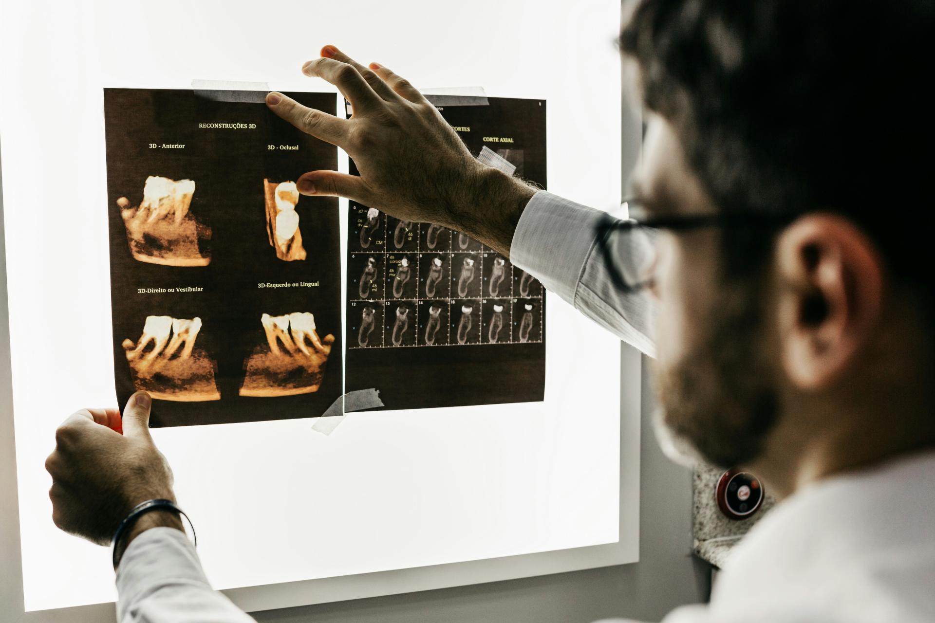
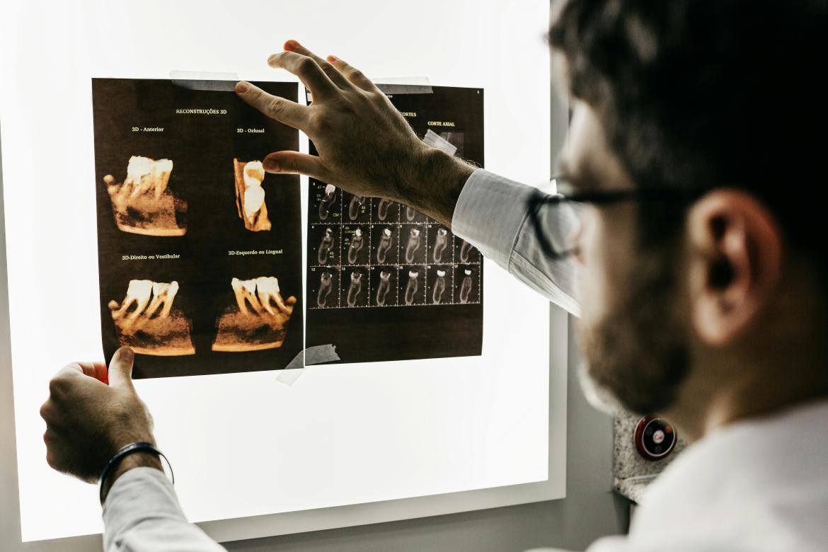
“The idea would be this — a patient comes to the dentist, and an X-ray image is taken for other reasons. For example, they are planning dental implants, and a 3D X-ray is taken, which is basically considered standard in modern implantology. But in addition to that, using this X-ray, the patient’s risk of osteoporosis is also determined,” the researcher explained. “That may perhaps sound a little strange, but it should be understood that jaw bones, which are part of our skeleton, are in the dentist’s field of view.”
She and her colleagues have been studying the idea that dentists, by examining jaw bones, could help diagnose osteoporosis for a long time — since her doctoral studies. “At first, we used panoramic radiographs and found that there are specific changes which, when identified, indicate quite accurately that a person may have an increased risk of osteoporosis. For the most part, we studied postmenopausal women, who are also a risk group, and the results were actually very good,” the researcher said.
At that time, 2D, or panoramic, X-rays were widely used in dental practices in Latvia, and the changes in jaw bones caused by osteoporosis could be clearly seen in them. However, quite soon a new technology appeared — 3D X-rays, which are now already much more popular.
Therefore, the studies had to be started anew to find out whether the same changes could also be seen in these three-dimensional images.
“We realised that, in fact, in this way we can see the bone structure even better — we can measure and assess even more accurately whether it indicates a risk of osteoporosis,” Slaidiņa revealed.
Dentists do not want to make such a serious diagnosis
In fact, researchers in other countries around the world have been discussing this method for many years, but in reality it has not really been implemented anywhere, because it would mean that dentists need additional training so that they are able to see and measure these changes in the jaw bones, which is not at all that simple. Moreover, many dentists do not feel sufficiently confident in this field to make such a serious diagnosis, and therefore indicate in various surveys conducted by researchers that they do not want to do so.
However, researchers in Latvia have now found a potential solution to this. “What can come to the rescue nowadays? Artificial intelligence! In principle, we want it to do the same thing that a dentist would do. We have a specific clinical protocol, and we want artificial intelligence to do exactly the same thing, and we hope that perhaps it will even do it better than a human, because, for example, it will never be tired,” the researcher said.
Previous studies show that the performance of artificial intelligence in recognising osteoporosis in X-rays taken by dentists is certainly no worse than that of a human. Researchers are currently trying to find different approaches to make the result even better.
At the same time, it is clear that the result must also be one that dentists are able to verify. “Of course, in medicine we do not want the artificial intelligence process to be placed in some kind of black box where we do not know what it is doing inside — a result comes out, but we do not know how it has produced it. That is why we are trying to control this system so that we understand how it performs each stage and so that each stage is carried out correctly,” Slaidiņa emphasised.
The aim — to detect the disease early
It is therefore important that the dentist, for example, is shown an image that clearly indicates where and what was measured, and which markers or maps the artificial intelligence worked with, so that the doctor can verify that this tool has done everything correctly.
“Of course, we do not claim to provide absolute diagnosis — it will be more of a screening method. We indicate that there is a risk, and after that the patient undergoes the gold-standard examination — osteodensitometry, where the diagnosis is confirmed or ruled out, and only then does some kind of treatment need to be started,” the researcher noted.
The main risk group for osteoporosis is postmenopausal women, while in men the risk increases a little later — after the age of 70. However, it would be most advisable for any person over the age of 65 to undergo osteodensitometry at least once.
Unfortunately, in real life this does not happen, and osteoporosis is often diagnosed only after the first fractures, which can be severe. It is possible that preventive assessment of the jaw bones at the dentist’s office would make it possible to detect this disease earlier, Slaidiņa said: “We would like this disease to be detected early and for patients to begin treatment early, before the first fracture has occurred, because in fact quite good medications are available nowadays, and they are also developing, changing and improving. As a result, osteoporosis can be quite successfully stopped and controlled, which makes timely diagnosis even more important.”